
Anatomy
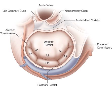
The mitral valve is a bileaflet valve comprised of anterior and posterior leaflets. It sits between the upper and lower chambers of the left side of the heart (between the left atrium and left ventricle)
It is a one-way valve which can become leaky (regurgitation) or narrowed (stenosis)
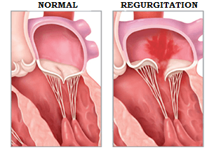
Mitral valve regurgitation
Primary (degenerative) leaks can involve one or both leaflets, and if amenable, repair is preferable
Secondary (functional) leaks involve the structures surrounding the leaflets rather than the leaflets themselves. Repair of this type of leak has be shown to be less durable. MitraClip has been shown to be beneficial in functional regurgitation. If a patient is not a candidate for MitraClip, then replacement of the mitral valve may be considered
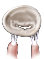
Mitral Stenosis
Narrowing of the mitral valve which reduces forward flow
Stenosis of the mitral valve is usually treated with valve replacement
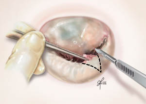
Mitral Repair
Using a variety of techniques to preserve the native valve and eliminate regurgitation

Mitral Replacement
The native valve leaflets are removed and a new valve (tissue or mechanical) is implanted

Tissue valves
Advantage: Avoids life-long anticoagulation (blood thinning)
Disadvantage: Will wear away with time
Mechanical valves
Advantage: Long-term durability
Disadvantage: Requires life-long anticoagulation (blood thinning)
